Microscopy
Microscopic examination of paint cross-sections
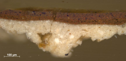
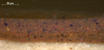
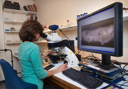
By examining minute samples of paint in cross-section under the microscope, the layer structure of a painting can be determined for that sample point. The ground layers are usually included in the sample. Many pigments can be identified by their optical properties in cross-sections, and analysis by scanning electron microscope with energy-dispersive X-ray (SEM-EDX) can be carried out on individual pigment particles and paint layers. Samples are mounted in blocks of cold-setting resin, then ground and polished to reveal the edge of the sample for examination in reflected (incident) light under the optical microscope. The usual magnification range is 60–1000X.
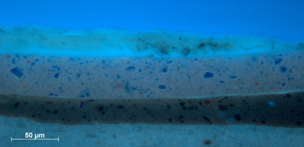
UV fluorescence microscopy
Some materials used in paintings fluoresce when exposed to ultraviolet radiation, that is they emit visible light of a specific wavelength (and therefore colour) when excited by the high energy ultraviolet. A light microscope is used to study properties of organic or inorganic substances using the phenomena of fluorescence in addition to reflection and absorption. When a paint cross-section is illuminated with ultraviolet light, certain materials may exhibit characteristic fluorescence.