SEM
Scanning electron microscopy (SEM)
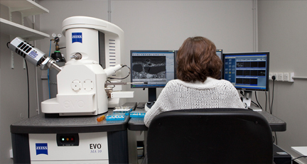
The scanning electron microscope is an instrument capable of revealing fine details of objects at magnifications far greater than is possible with the optical (light) microscope. The SEM uses a focused beam of electrons to scan the surface of a sample under examination, generating several types of signal each collected by a different type of detector. The signals of particular interest for the purposes of paint analysis are: secondary electrons (SE); backscatter electrons (BSE); and characteristic X-rays.
Secondary electrons (SE) interact with surfaces in a particular way allowing very detailed topography and three-dimensional structure of a micro-sample of paint to be imaged and recorded. Thus very high-resolution images can be produced of a sample surface at magnifications of up to 100,000x. However, because it is not the product of visible light, there is no colour in an SEM image (SEM-micrograph).
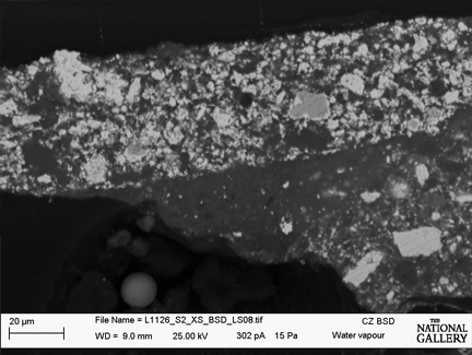
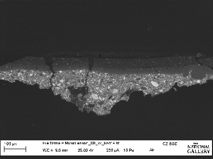
The intensity of the backscatted electron signal is strongly related to the atomic number of the specimen, and so BSE images can help provide information about the distribution of different elements in the sample. For example, an element of high atomic number, such as lead (Pb), will appear very bright in a BSE image, while an element of lower atomic number such as calcium (Ca) will appear much darker. These images are often used in conjunction with the spectra and maps made from the characteristic X-rays.
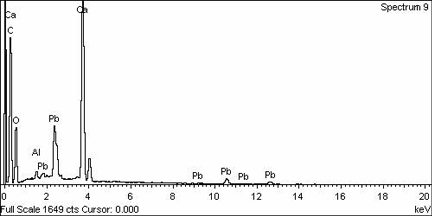
Energy dispersive X-ray spectrometer / Energy-dispersive X-ray microanalysis (EDS or EDX)
This technique is a method of elemental analysis carried out in the scanning electron microscope (SEM). Small areas of a sample which has been imaged in the SEM can be selected and analysed for their constituent elements. For example, a single paint layer in a cross-section, or an individual pigment particle of very small size, can be analysed selectively. The electron beam which falls on the specimen in the SEM generates small amounts of X-rays which are characteristic in their energies of the elements in the sample that produce them. The instrument (spectrometer) measures the energies of these X-rays and assigns them on a visual display to the elements present. For the purposes of pigment identification in paint samples, the results show whether the sample contains, for example, the chemical elements lead, copper, arsenic, tin, antimony, cobalt, chromium and so on, but not what the actual component pigments are. These analytical data must then be interpreted in conjunction with examination of paint samples by optical microscopy in order to determine which pigments are actually present. Modern forms of these instruments allow the generation of 'element maps' in such a way that the distribution of chemical elements, for example in a paint cross-section, can be visualised and recorded.